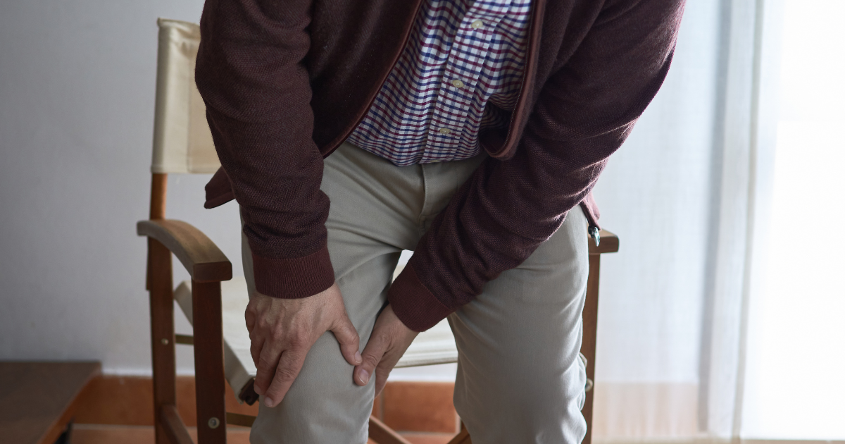

Gout is a type of inflammatory arthritis that develops when uric acid forms crystals in and around the joints. These crystals can trigger sudden episodes of severe pain, swelling, redness, and tenderness. Although gout often affects the big toe, it can also involve the ankles, knees, wrists, fingers, and elbows.
Many people seek medical attention only after experiencing repeated gout attacks. However, uric acid crystals may accumulate in the joints and surrounding tissues even before noticeable symptoms develop. If gout remains uncontrolled for years, it can lead to tophi, chronic inflammation, and permanent joint damage.
An important question is whether gout can be detected before these complications occur. Musculoskeletal ultrasound is a useful imaging technique that can help identify urate crystal deposits and inflammation associated with gout. But how early can it detect the disease, and can it predict future joint damage?
Let’s understand how ultrasound helps doctors diagnose and manage gout.
What Is Musculoskeletal Ultrasound?
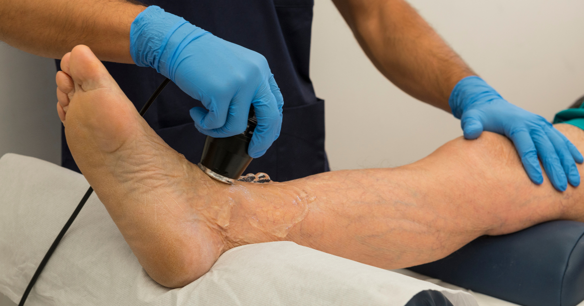
Musculoskeletal ultrasound is a non-invasive imaging test that uses high-frequency sound waves to produce images of joints, tendons, ligaments, and surrounding soft tissues.
Unlike X-rays, ultrasound does not use ionizing radiation. It allows doctors to examine joints in real time and look for signs of inflammation or crystal deposits.
For patients with suspected gout, ultrasound can help identify:
- Uric acid crystal deposits in and around joints.
- Tophi, which are collections of urate crystals in soft tissues.
- Inflammation of the joint lining.
- Fluid accumulation inside a joint.
- Inflammation around tendons and other soft tissues.
- Certain changes associated with long-standing gout.
The examination may focus on the joint causing symptoms, such as the big toe, ankle, or knee. Depending on the clinical situation, doctors may also examine other joints where gout-related deposits commonly occur.
Can Ultrasound Detect Gout Before Joint Damage Begins?
Yes, ultrasound can sometimes detect urate crystal deposits before permanent joint damage becomes visible on conventional X-rays.
Gout develops when monosodium urate crystals accumulate because uric acid levels have remained high enough over time. These crystals may collect on the surface of cartilage, inside joints, and around tendons.
Ultrasound can reveal characteristic changes associated with these deposits. In some patients, it can identify evidence of crystal accumulation even when symptoms are mild or when there has been no obvious structural damage.
However, ultrasound cannot guarantee that gout will be detected at the earliest possible stage. Small deposits may be difficult to identify, and a normal scan does not completely rule out gout.
It is also important to understand that high uric acid alone does not mean a person has gout. Some people have elevated uric acid levels for years without developing crystal-related arthritis. Ultrasound findings must therefore be interpreted alongside symptoms, examination findings, blood tests, and medical history.
What Can Ultrasound Reveal in Gout?
Doctors look for several characteristic findings when performing an ultrasound examination for suspected gout.
1. The Double-Contour Sign
The double-contour sign is one of the characteristic ultrasound findings associated with gout.
Normally, joint cartilage has a smooth appearance on ultrasound. In gout, urate crystals may accumulate on the surface of the cartilage, creating an additional bright line that appears parallel to the underlying bone surface.
This appearance is called the double-contour sign.
It can help doctors identify urate deposition, sometimes before conventional X-rays show gout-related bone damage.
However, this sign is not present in every person with gout, and similar appearances can occasionally occur in other conditions. The finding must be interpreted in its clinical context.
2. Tophi or Urate Crystal Deposits
Tophi are collections of monosodium urate crystals that develop in soft tissues.
They may appear as lumps under the skin, particularly around the fingers, toes, elbows, and ears. Some are visible or painful, while others remain unnoticed for a long time.
Ultrasound can help identify tophi beneath the skin or within tissues around joints. It can also help assess their size and location.
Detecting tophi may be useful because their presence can indicate a substantial burden of urate crystals and may influence decisions about long-term uric acid-lowering treatment.
3. Joint Inflammation
During a gout flare, inflammation can cause swelling, pain, redness, and warmth around an affected joint.
Ultrasound may reveal fluid within the joint, thickening of the joint lining, and increased blood flow associated with inflammation when Doppler imaging is used.
These findings can support the assessment of an active inflammatory process. However, they are not specific to gout and may also occur in infections and other forms of arthritis.
4. Tendon and Soft-Tissue Involvement
Gout does not affect only the joint itself. Urate crystals can also accumulate around tendons and other nearby soft tissues.
Ultrasound may identify deposits around tendons, including those near the ankle and elbow.
This can be helpful when a patient has persistent swelling or pain in an area where gout is suspected but the symptoms do not resemble a typical big-toe attack.
Can Ultrasound Detect Gout Before Symptoms Appear?
In some cases, ultrasound can identify urate crystal deposits in people who have never experienced a typical gout attack.
This is important because crystal deposition and symptomatic gout are not exactly the same thing.
A person may have elevated uric acid levels and detectable crystal deposits without experiencing pain or swelling. This does not automatically mean that the person has symptomatic gout or requires medication.
Doctors consider several factors when deciding what these findings mean, including:
- Whether the patient has experienced previous gout attacks.
- The blood uric acid level and overall medical history.
- Whether characteristic crystal deposits or tophi are present.
- Kidney function and other relevant health conditions.
- The presence of other risk factors or possible explanations for symptoms.
Routine ultrasound screening is not generally necessary for every person with elevated uric acid but no symptoms. Imaging is most useful when it answers a specific clinical question.
Can Ultrasound Predict Future Joint Damage?
Ultrasound can identify crystal deposits and some signs of inflammation that may not be visible on an ordinary X-ray. These findings can help doctors understand the extent of gout-related changes.
However, ultrasound cannot accurately predict exactly when or whether an individual will develop permanent joint damage.
The risk of complications depends on several factors, including the duration of gout, the amount of crystal accumulation, the frequency of inflammatory attacks, and how effectively uric acid levels are controlled.
When gout remains untreated or poorly controlled, crystals may continue to accumulate. Over time, some patients develop persistent inflammation, tophi, and bone erosions.
Early recognition and appropriate treatment can help reduce this risk.
The goal is not simply to find crystals on a scan. It is to determine whether the findings indicate gout and whether treatment or closer monitoring is appropriate.
Ultrasound vs X-Ray vs MRI: Which Is Better for Gout?
Different imaging tests provide different types of information.
Ultrasound
Ultrasound is particularly useful for identifying characteristic urate deposits, tophi, joint fluid, and soft-tissue inflammation.
Advantages:
- Does not use ionizing radiation.
- Can be performed at the bedside or in an outpatient clinic.
- Allows real-time assessment of joints and surrounding tissues.
- Can help guide a joint aspiration when needed.
Limitations:
- Results depend on the operator’s experience.
- Small or early deposits may be missed.
- Some findings overlap with those of other diseases.
- It cannot reliably exclude gout when the scan is normal.
X-Ray
X-rays can reveal certain bone changes associated with long-standing gout, including characteristic erosions.
However, early gout may not produce visible changes on an X-ray. This makes X-rays less useful for identifying early crystal deposition.
They can still be valuable when doctors need to assess established bone damage or investigate other causes of joint pain.
MRI
Magnetic resonance imaging, or MRI, provides detailed images of soft tissues, bone marrow, joints, and surrounding structures.
It may be useful when doctors suspect complications or need to investigate another condition. However, MRI is not usually the first imaging choice for identifying urate crystals because its findings are generally less specific for gout than characteristic ultrasound or dual-energy CT findings.
Dual-Energy CT
Dual-energy computed tomography is a specialized imaging technique that can help identify and map urate deposits.
It may be useful in selected patients when the diagnosis remains uncertain or when doctors need additional information about crystal deposits.
The best test depends on the patient’s symptoms, clinical examination, medical history, and the specific question the doctor needs to answer.
Can a Blood Test for Uric Acid Replace Ultrasound?
No. Blood tests and ultrasound provide different information.
A serum uric acid test measures the amount of uric acid in the blood. It helps doctors assess risk and monitor treatment, but it cannot independently confirm or exclude gout.
Some people have high uric acid without gout. Conversely, uric acid levels can fall into the normal range during an acute gout attack.
Ultrasound may provide evidence of crystal deposition, while a blood test measures circulating uric acid.
When the diagnosis is uncertain, doctors may recommend additional investigations. Examining fluid collected from an affected joint for monosodium urate crystals remains an important diagnostic method when it can be performed.
A correct diagnosis matters because several conditions, including septic arthritis and other inflammatory joint diseases, can cause symptoms similar to gout.
Who May Benefit From an Ultrasound for Gout?
A doctor may consider musculoskeletal ultrasound in the following situations:
- Uncertain diagnosis: Symptoms suggest gout, but the diagnosis is not clear.
- Repeated attacks: The patient experiences recurrent episodes of joint pain and swelling.
- Possible tophi: There are unexplained lumps around joints or tendons.
- Atypical symptoms: Pain affects less typical areas, such as the wrist, fingers, or tendons.
- Suspected crystal deposits: The doctor needs additional evidence of urate accumulation.
- Treatment assessment: Imaging may help evaluate crystal deposits in selected patients when the result could influence management.
Ultrasound is not necessary for every person with gout. A doctor determines whether it will provide useful information beyond the patient’s symptoms, examination, and laboratory results.
Can Early Gout Detection Prevent Permanent Joint Damage?
Early diagnosis can help patients begin appropriate treatment before recurrent inflammation and crystal accumulation cause serious complications.
For people with confirmed gout, long-term management often includes uric acid-lowering medication when indicated, regular monitoring, and lifestyle measures that support overall health.
For most patients receiving urate-lowering treatment, a common target is a serum uric acid level below 6 mg/dL. Some patients with severe gout or substantial tophi may need a lower target, depending on their clinical circumstances and the treatment approach recommended by their doctor.
Medication should not be stopped or changed without medical advice, even when pain disappears. Controlling uric acid over time helps dissolve existing crystals and reduce the likelihood of future attacks.
Lifestyle measures, such as maintaining adequate hydration when medically appropriate, limiting alcohol and sugary drinks, and achieving a healthy weight gradually, can support treatment. However, lifestyle changes alone may not be sufficient for people with established gout.
The key is to diagnose gout correctly and control uric acid effectively—not simply to rely on an imaging test.
Frequently Asked Questions About Ultrasound for Gout
1. Can ultrasound detect gout in its early stages?
Yes. Ultrasound can sometimes identify characteristic urate crystal deposits before conventional X-rays show bone damage. However, early deposits may be missed, so a normal scan does not rule out gout.
2. Can ultrasound detect gout when uric acid levels are normal?
Ultrasound may show evidence of urate deposits even when the blood uric acid level is normal at the time of testing. The findings must be interpreted alongside symptoms and other clinical information.
3. Is ultrasound better than an X-ray for diagnosing early gout?
Ultrasound is generally more useful for detecting characteristic crystal deposits and soft-tissue changes, whereas X-rays are more useful for assessing certain structural changes associated with long-standing gout.
4. Can ultrasound show uric acid crystals directly?
Ultrasound does not show individual crystals in the same way that a microscope can. Instead, it identifies characteristic appearances associated with urate deposits, such as the double-contour sign and tophi.
5. Does everyone with high uric acid need an ultrasound?
No. Routine ultrasound is not generally recommended for everyone with elevated uric acid but no symptoms. A doctor may suggest imaging when there is a specific diagnostic or management question.
6. Can ultrasound tell whether gout has permanently damaged a joint?
Ultrasound can identify certain changes associated with gout, but it cannot reliably assess every type of permanent bone or joint damage. X-rays, CT, MRI, or other investigations may be appropriate depending on the clinical situation.
7. Should I get an ultrasound if I have repeated gout attacks?
If you experience repeated attacks, consult your doctor to review the diagnosis, uric acid levels, and treatment plan. Ultrasound may be useful if the diagnosis is uncertain or if identifying crystal deposits would change management.
Conclusion
Musculoskeletal ultrasound is a valuable tool in the assessment of gout. It can reveal characteristic urate deposits, tophi, and inflammation, sometimes before conventional X-rays show structural damage.
However, ultrasound is not a standalone test, and a normal result does not completely exclude gout. Diagnosis requires a careful assessment of symptoms, examination findings, blood tests, and, when necessary, joint fluid analysis or other imaging.
If you experience repeated episodes of joint pain, swelling, or redness, seek medical evaluation rather than relying only on a uric acid blood test. Recognizing gout early and maintaining appropriate uric acid control can help prevent recurrent attacks and reduce the risk of long-term joint damage.